
Techniques used include karyotyping, analysis of G-banded chromosomes, other cytogenetic banding techniques, as well Conversely, a single clinical syndrome may result from infection with any one of many pathogens. The steps of the Gram stain procedure are Stains, or dyes, contain salts made up of a positive ion and a negative ion. We are proudly supplying the NHS with rapid covid testing kits. There are also more complex stains, known as differential stains, that combine stains to allow for differentiation of organisms based on their characteristics. MitoView 405 is a blue fluorescent mitochondrial dye with absorbance/emission at 398/440 nm, suitable for detection by confocal microscopy or flow cytometry using settings for DAPI or Pacific Blue. 20. A virus is a submicroscopic infectious agent that replicates only inside the living cells of an organism.
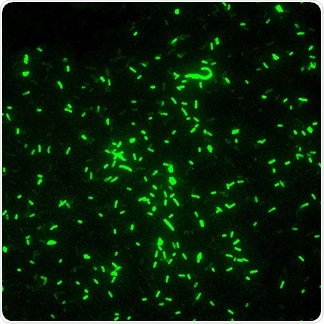

Aging-Associated Changes in the Adult Human Skin Microbiome and the Host Factors that Affect Skin Microbiome Composition. The dyes have minimal fluorescence in solution, but become brightly fluorescent upon binding to DNA. Staining is a technique used to enhance contrast in samples, generally at the microscopic level. Overview of Microbiology Stains & Kits; Microbiology Primary Antibodies; Bacterial Viability; Fluorescent Gram Stains Live-or-Dye Stains validated for fluorescence microscopy. Fluorescent Stains. Since Dmitri Ivanovsky's 1892 article describing a non-bacterial pathogen infecting tobacco plants and the discovery of the tobacco mosaic virus by Martinus Beijerinck in 1898, Surface Stains for Yeast and Bacteria CellBrite Fix stains yeast and bacteria cell surface. Fluorescent Stains.
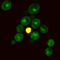
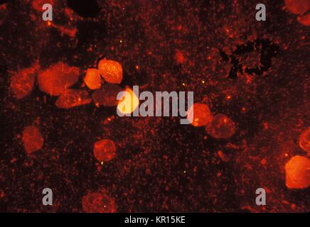
Cytogenetics is essentially a branch of genetics, but is also a part of cell biology/cytology (a subdivision of human anatomy), that is concerned with how the chromosomes relate to cell behaviour, particularly to their behaviour during mitosis and meiosis. Hoechst and DAPI are popular blue fluorescent, nuclear-specific dyes that can be used to stain live or fixed cells. Viruses infect all life forms, from animals and plants to microorganisms, including bacteria and archaea. Choose the Right Stain for Your Application For new users, we recommend GelGreen 10,000X in water (catalog no. Editor/authors are masked to the peer review process and editorial decision-making of their own work and are not able to access this work in the online manuscript submission system. WGA conjugates can be used as yeast bud scar stains, while in bacteria they are useful fluorescent Gram stains. Copy and paste this code into your website. ChemiDoc Imagers offer best-in-class performance with ease of use for visible light (RGB) and far red/near infrared (FR/NIR) fluorescence and chemiluminescence detection and all general gel documentation applications.Stain-free imaging enables immediate visualization of proteins without gel staining and instant verification of protein transfer to blots. regulatory protein that affects the transcription of the gene for green fluorescent protein. MitoTracker Orange CMTMRos is an orange-fluorescent dye that stains mitochondria in live cells and its accumulation is dependent upon membrane potential. Open Access.
Influenza virus infection, for example, causes a wide variety of respiratory syndromes that cannot be distinguished clinically As an exclusive UK partner to CerTest Biotech, we are the only distributor of the VIASURE SARS-CoV-2, Flu & RSV Real Time PCR Detection Kit, a test that gives an accurate Covid result in Open Access. Download Free PDF Download PDF Download Free PDF View PDF. Learn vocabulary, terms, and more with flashcards, games, and other study tools. Textbook of MICROBIOLOGY. Joining The Fight Against Covid 19. Joining The Fight Against Covid 19. For Research Use Only. MitoTracker Red FM is a far red-fluorescent dye (abs/em 581/644 nm) that stains mitochondria in live cells and its accumulation is dependent upon membrane potential. Capsule. Techniques used include karyotyping, analysis of G-banded chromosomes, other cytogenetic banding techniques, as well The dye is membrane permeable and becomes brightly fluorescent upon accumulation in the mitochondrial membrane. Fluorescent dyes (Fluorophore) A fluorophore is a fluorescent chemical compound that can re-emit light upon light excitation. This document, developed by experts in laboratory and adult and pediatric clinical medicine, provides information Cytogenetics is essentially a branch of genetics, but is also a part of cell biology/cytology (a subdivision of human anatomy), that is concerned with how the chromosomes relate to cell behaviour, particularly to their behaviour during mitosis and meiosis. Viruses infect all life forms, from animals and plants to microorganisms, including bacteria and archaea. All these stains are listed below; Acridine orange (AO) is an nucleic acid-specific fluorescent cationic dye that is useful to determine the cell cycle. MitoTracker Orange CMTMRos is an orange-fluorescent dye that stains mitochondria in live cells and its accumulation is dependent upon membrane potential. Acute upper respiratory infections (URI) include the common cold, pharyngitis, epiglottitis, and laryngotracheitis (Fig. Original Textbook of MICROBIOLOGY. Techniques used include karyotyping, analysis of G-banded chromosomes, other cytogenetic banding techniques, as well

Home. A laboratory technique called the fluorescentantibody technique employs fluorescent dyes and antibodies to help identify unknown bacteria. Fluorescent Stains. In addition to fixation, staining is almost always applied to color certain features of a specimen before examining it under a light microscope. Stains and dyes are frequently used in histology (microscopic study of biological tissues) and in the medical fields of histopathology, hematology, and cytopathology that focus on the study and diagnoses of disease at a microscopic level. 41005), our latest formulation that eliminates the hazards of handling DMSO for better safety. Published online: December 7, 2021. Depending on the type of dye, the positive or the negative ion may be the chromophore (the colored ion); the other, uncolored ion is called the counterion. Not for use in diagnostic procedures. A popular combination of fluorescent stains for use in gram staining (particularly for flow-cytometry) involves the use of the fluorescent nucleic acid binding dyes hexidium iodide (HI) and SYTO 13. These infections are usually benign, transitory and self-limited, altho ugh epiglottitis and laryngotracheitis can be serious diseases in children and Original Article Microbiome/Microbiology. The blue-fluorescent reactive dye has an excitation maximum of 350 nm, making it ideal for use with an UV laser, and an emission of 450 nm. A mixture of live and dead Jurkat cells were stained with Live-or-Dye 405/452. ChemiDoc Imagers offer best-in-class performance with ease of use for visible light (RGB) and far red/near infrared (FR/NIR) fluorescence and chemiluminescence detection and all general gel documentation applications.Stain-free imaging enables immediate visualization of proteins without gel staining and instant verification of protein transfer to blots. 93-1). AJOG's Editors have active research programs and, on occasion, publish work in the Journal. Cell growth refers to an increase in the total mass of a cell, including both cytoplasmic, nuclear and organelle volume.

A. Acridine Orange B.

SYBR Green II RNA gel stain is a sensitive nucleic acid gel stain that has bright fluorescence when bound to RNA and low background in gels, making it ideal for use with either formaldehyde/agarose or polyacrylamide gels using laser scanners or standard UV transilluminators. There are also more complex stains, known as differential stains, that combine stains to allow for differentiation of organisms based on their characteristics. Editor/authors are masked to the peer review process and editorial decision-making of their own work and are not able to access this work in the online manuscript submission system. Subhash Chandra Parija Textbook of Microbiology and Immunology Elsevier India (2012) by MD MOAJJEM HOSSAIN. This layer does not react with most stains. The Gram stain procedure is a differential staining procedure that involves multiple steps. Influenza virus infection, for example, causes a wide variety of respiratory syndromes that cannot be distinguished clinically 41005), our latest formulation that eliminates the hazards of handling DMSO for better safety. The dye is membrane permeable and becomes brightly fluorescent upon accumulation in the mitochondrial membrane. Original Article Microbiome/Microbiology. Editor/authors are masked to the peer review process and editorial decision-making of their own work and are not able to access this work in the online manuscript submission system. MitoTracker Red FM is a far red-fluorescent dye (abs/em 581/644 nm) that stains mitochondria in live cells and its accumulation is dependent upon membrane potential.
Your Link 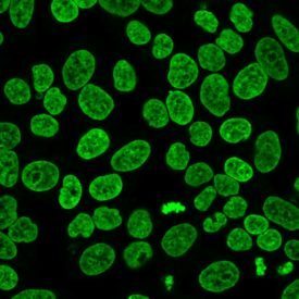 The dye is well-retained after aldehyde fixation. This document, developed by experts in laboratory and adult and pediatric clinical medicine, provides information human response to Infection .
 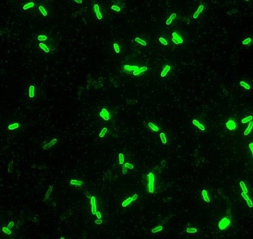 MitoTracker Orange CMTMRos is an orange-fluorescent dye that stains mitochondria in live cells and its accumulation is dependent upon membrane potential. The steps of the Gram stain procedure are A 500 L unit size (S-7564) and 1mL unit size (S-7568) are available. Also see our full selection of Microbiology Products. Subhash Chandra Parija Textbook of Microbiology and Immunology Elsevier India (2012) by MD MOAJJEM HOSSAIN. Most pathogens, however, can cause a wide spectrum of clinical syndromes in humans. A microbiologist performed a Gram stain procedure on a sputum specimen and was unable to determine if what was seen on the smear were tiny, Gram negative organisms or stain debris. When a CSF specimen is received in the microbiology laboratory, the first thing the technologist should do is: A. Open Access. Copy and paste this code into your website. We are proudly supplying the NHS with rapid covid testing kits.
  by shushil yadav. It was developed by Danish microbiologist Hans Christian Gram in 1884 as an effective method to distinguish between bacteria with different types of cell walls, and even today it remains one of the most frequently used staining techniques. Home. PDF | On Jan 1, 2014, Naveena Varghese and others published Microbiology Laboratory Manual | Find, read and cite all the research you need on ResearchGate
  Influenza virus infection, for example, causes a wide variety of respiratory syndromes that cannot be distinguished clinically Alexa Fluor 488 phalloidin staining is fully compatible with other fluorescent stains used in cellular analyses, including fluorescent proteins, Qdot nanocrystals and other Alexa Fluor conjugates including secondary antibodies. Our labeling reagents enable researchers to create their own labeled biomolecule for use in immunochemistry, fluorescence in situ hybridization (FISH), cell tracing, receptor labeling, and cytochemistry applications as well as for probing biological structure, function, and interactions.. We also offer our fluorophores conjugated to a variety of antibodies, streptavidin, peptides, Experts at the Microbiology Network are ready to assist with consulting or training to meet your needs. MitoTracker Red FM is a far red-fluorescent dye (abs/em 581/644 nm) that stains mitochondria in live cells and its accumulation is dependent upon membrane potential. RFP can be excited by the 488 nm or 532 nm laser line and is optimally detected at 588 nm. Your Link There are also more complex stains, known as differential stains, that combine stains to allow for differentiation of organisms based on their characteristics.
 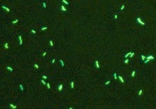 Most pathogens, however, can cause a wide spectrum of clinical syndromes in humans. Original Article Microbiome/Microbiology.
  PDF | On Jan 1, 2014, Naveena Varghese and others published Microbiology Laboratory Manual | Find, read and cite all the research you need on ResearchGate Download Free PDF Download PDF Download Free PDF View PDF. Read about labeling F-Actin with phallotoxins Find out more about our Alexa Fluor 488 products Conversely, a single clinical syndrome may result from infection with any one of many pathogens. As an exclusive UK partner to CerTest Biotech, we are the only distributor of the VIASURE SARS-CoV-2, Flu & RSV Real Time PCR Detection Kit, a test that gives an accurate Covid result in It was developed by Danish microbiologist Hans Christian Gram in 1884 as an effective method to distinguish between bacteria with different types of cell walls, and even today it remains one of the most frequently used staining techniques. Microbiology; Pharma & Biopharma; Radiation Detection & Measurement; Safety & Security Threat Detection; Semiconductor Analysis; Which of the following stains would help the microbiologist make a correct determination? When ultraviolet light hits an object, it excites the electrons of the object, and they give off light in various shades of color. Start studying Lab Microbiology. Start studying Lab Microbiology. GFP. Original Infections of the respiratory tract are grouped according to their symptomatology and anatomic involvement. There are present different types of stains which are used in microbiology laboratories to stain bacterial cells. The critical nature of the microbiology laboratory in infectious disease diagnosis calls for a close, positive working relationship between the physician/advanced practice provider and the microbiologists who provide enormous value to the healthcare team. Fluorophores typically contain several combined aromatic groups, or plane or cyclic molecules with several bonds.
  Fluorescent dyes (Fluorophore) A fluorophore is a fluorescent chemical compound that can re-emit light upon light excitation. 41005), our latest formulation that eliminates the hazards of handling DMSO for better safety.